July 22, 2026

The InnoRenew CoE research laboratories include a laboratory for microscopy in which structures and morphology of renewable materials are analysed.
The laboratory has equipment for the preparation of samples that will be observed under microscopes, the microscopes themselves, as well as a variety of other devices for characterisation of physical properties.
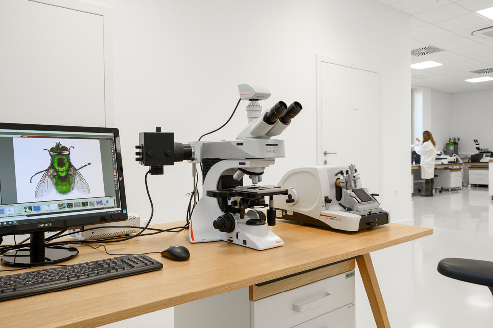
Image: Miran Kambič
Preparation of our samples typically begins with larger pieces that we cut in our wood workshop on standard table saws. We then take these smaller sections into the lab and use a detailed surface preparation device that can isolate small regions of interest to refine using milling heads and polishing wheels. This results in highly polished surface that to the human eye is mirror like. This can further be refined using our ion beam mill. This device, akin to a sand blaster, creates a near perfect surface for microscopic analysis. This is used for specimens that are otherwise impossible to cut using conventional blades that can damage the internal structure. Another approach to tissues, like wood, is to take thin sections of the material for standard light microscopy. This approach softens the wood, slices the specimens into 10-30 micron thick sections, and then stains the specimens for better contrast and observation.
For the observation itself, we have several microscopes at our disposal including a fluorescent microscope, a 3D digital microscope, a surface metrology microscope, and a fluorescent microscope that is used for monitoring active growth of cultures. Most of these devices are automatised and can be programmed to capture large regions of interest at high resolution independently. This enables us to use detailed Z stacking across the entire surface of the specimens to return sharp images of surfaces that are rough and would otherwise have non-focused regions. This array of microscopes lets us analyse the internal structure, morphology, surface roughness and profile, identify marked components or regions of interest, and monitor development of living cultures. We can observe structures from microns to millimetres in this laboratory.
Beyond the microscopes this lab also houses some specialised equipment for characterisation of materials. This includes the measurement of surface energy, true density of materials, gas chromatography, size exclusion chromatography, and viscosity measurements. These devices complement the microscopy work by measuring important physical and chemical parameters used in material development and modification.
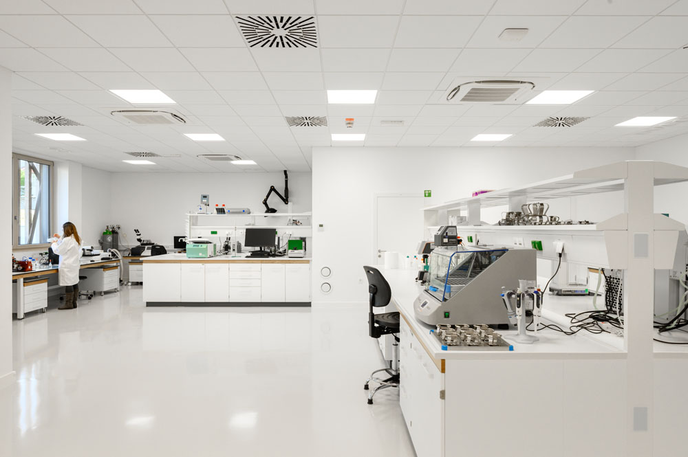
Image: Miran Kambič
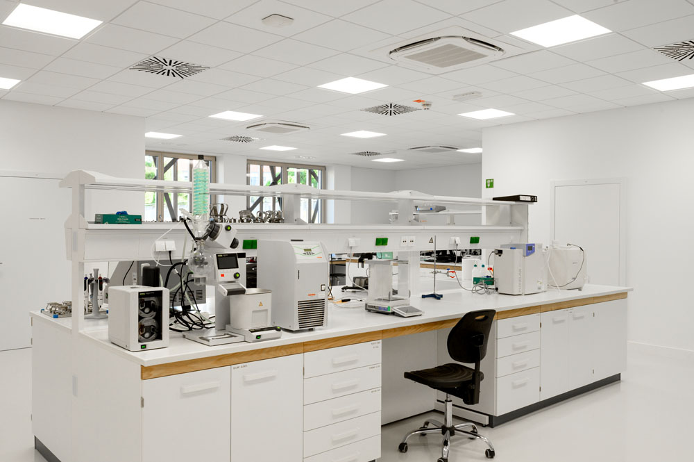
Image: Miran Kambič
The research being done in this laboratory ranges from simple identification of wood species to a strong physical/chemical analysis of modifications or treatments made on natural materials. This can support the industry when preparing new treatments, composite products, investigation of physical phenomena, analysis of volatile emissions from materials in buildings, development of new adhesive and coatings, etc. This lab provides fundamental analysis of materials at a microscopic level. Wood, other natural materials, and their constitutive components are highly structured and complicated natural materials that require careful consideration of how we affect their microscopic structure as this affects the rest of their performance in service.
List of equipment in the microscopy laboratory
Dr. Matthew Schwarzkopf, InnoRenew CoE